Skip to content
Living With Glioblastoma
My Life But Not as I Planned It
My Story
What is a Glioblastoma?
My Blog
Timeline and Treatment Info
Scans
Contact Me
Living With Glioblastoma
My Life But Not as I Planned It
Contact Me
My Blog
My Story
Scans
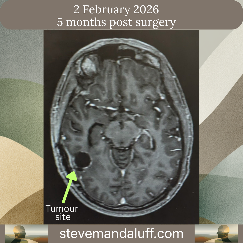
Scan 1: 2 February 2026
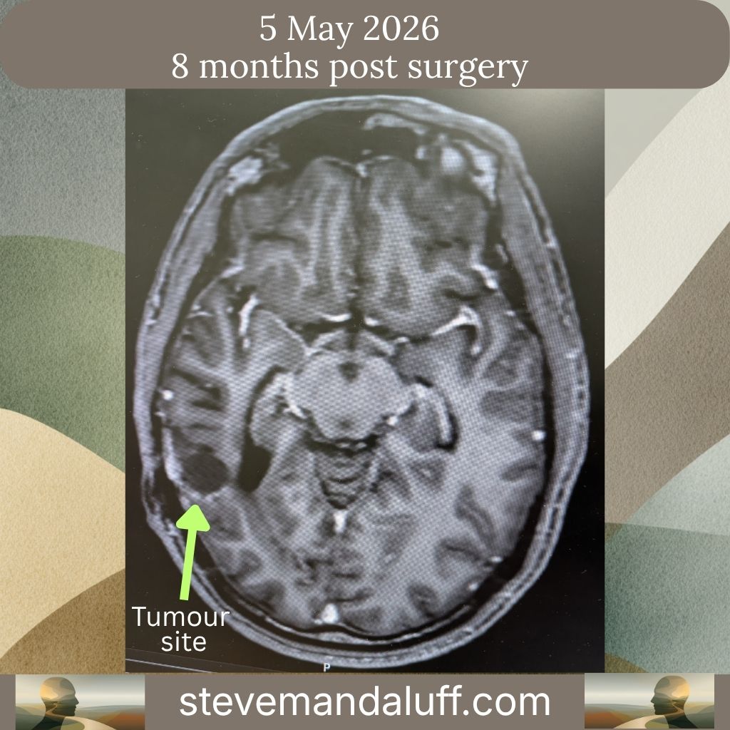
Scan 2: 5 May 2026
Timeline and Treatment Info
Welcome
What is a Glioblastoma?
Scans
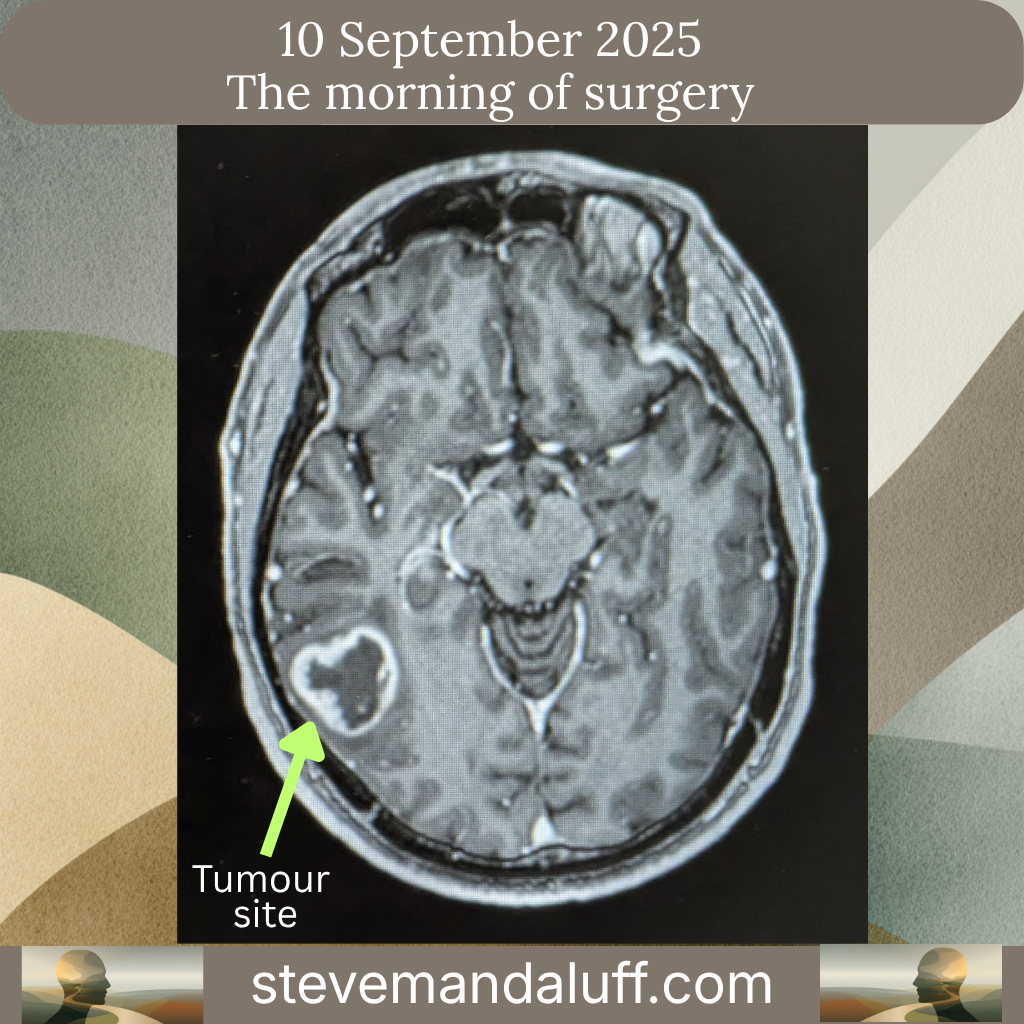
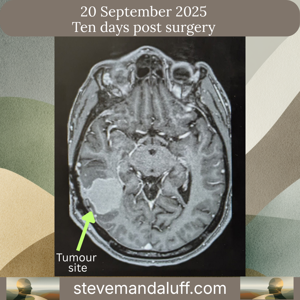
An overview of my scans so far:
Scan Details
Scan 1: 2 February 2026
Scan 2: 5 May 2026
Scroll to Top