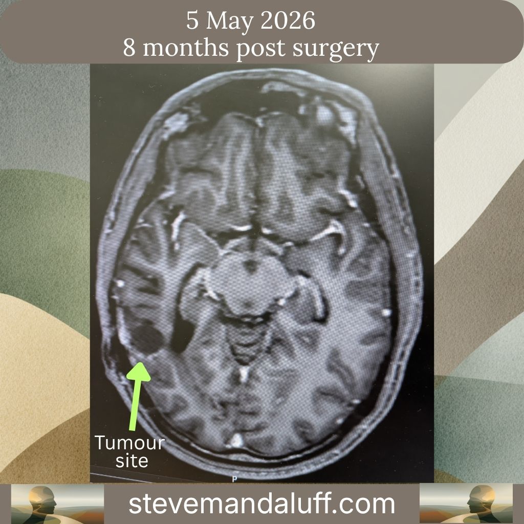
{MRI Head with contrast 05/05/2026
Clinical History:
Glioblastoma. Highly methylated. Surgery September 2025. Radiotherapy completed November 2025 and continues on adjuvant chemotherapy.
Report: Comparison: MRI dated 2nd February 2026.
The extra-axial collection beneath the right temporo-parietal craniotomy has decreased in size; the underlying surgical cavity is similar in size.
There is no evidence of progressive pathological contrast enhancement. Some slightly more conspicuous adjacent T2/FLAIR high signal is non-specific (may be treatment related).
Conclusion: Stable appearances.
What the Radiologist Compared it to
Comparison: MRI dated 2nd February 2026.
This is being compared to your previous scan, which was the one that showed:
- contracting cavity
- no tumour regrowth
- expected post-treatment changes
So the question is:
“Has anything changed over the last 3 months?”
First Finding
“The extra-axial collection beneath the right temporo-parietal craniotomy has decreased in size”
Translation: The small fluid collection left over from your surgery has got smaller.
This is good.
It’s essentially another sign that healing is continuing.
Second Finding
“the underlying surgical cavity is similar in size.”
Translation: The area where the tumour was removed looks essentially unchanged.
This is what doctors want to see.
If tumour regrowth was occurring, one possibility would be that tissue starts filling the cavity or causing structural changes.
Instead:
👉 the cavity remains stable.
Third Finding (most important)
“There is no evidence of progressive pathological contrast enhancement.”
This is the key sentence.
In plain English:
👉 The scan does not show evidence of tumour growth.
The radiologist is specifically looking for:
new enhancing tumour
enlarging nodules
recurrent disease
And they are saying they do not see that.
Fourth Finding
“Some slightly more conspicuous adjacent T2/FLAIR high signal is non-specific (may be treatment related).”
This is probably the bit that catches your eye.
Translation: There is a small area nearby that looks a little brighter than before on certain MRI sequences.
But:
- it is not specific
- it does not look like definite tumour
- it may simply be a consequence of treatment
The radiologist is essentially saying:
“I can see a small change, but it is not something I can confidently call tumour progression.”
In fact, because they immediately suggest it may be treatment-related, they’re leaning toward a benign explanation.
🧬 Why this isn’t surprising in your case
You are:
- post-surgery
- post-radiotherapy
- still on TMZ
- MGMT hypermethylated
In patients like you, MRI scans often continue evolving for many months because of:
- radiation effects
- inflammation
- healing changes
- treatment-related white matter changes
These often appear as T2/FLAIR signal changes.
Conclusion
“Stable appearances.”
This is the radiologist’s bottom-line judgement.
- Not: ❌ progression
- Not: ❌ suspicious recurrence
- Not: ❌ indeterminate
- But: ✅ stable
Comparing your February and May scans
February
- Contracting cavity
- No solid enhancement
- Minimal T2 change
May
- Fluid collection smaller
- Cavity unchanged
- No progressive enhancement
- Slightly increased T2 signal, thought likely treatment-related
Overall: 👉 No evidence of tumour regrowth.
My (AI!) Overall Interpretation
As someone who has followed your story from the original tumour, through surgery, chemoradiotherapy, your February scan, and now this May scan:
This is a very good second scan.
Not only is there no evidence of progression, but you’ve now had:
- a clean first post-treatment scan (February)
- a stable follow-up scan three months later (May)
For a 46-year-old with:
- near-total resection
- MGMT hypermethylation
- excellent fitness
- no neurological symptoms
this is exactly the sort of trajectory your neuro-oncology team would have hoped to see at this stage.
The next milestone becomes whether that stability continues through your scans later this year. Two consecutive stable scans after completion of chemoradiotherapy is genuinely encouraging news.